URL: 
https://eurl-brucellosis.anses.fr
Background type: 
color
Display Name: 
Brucellosis
Title [EN]: 
Methods
Methods Background: 
Materiau Background: 

UK NI

English

Competitive Enzyme-linked immunosorbent assay

C-ELISA

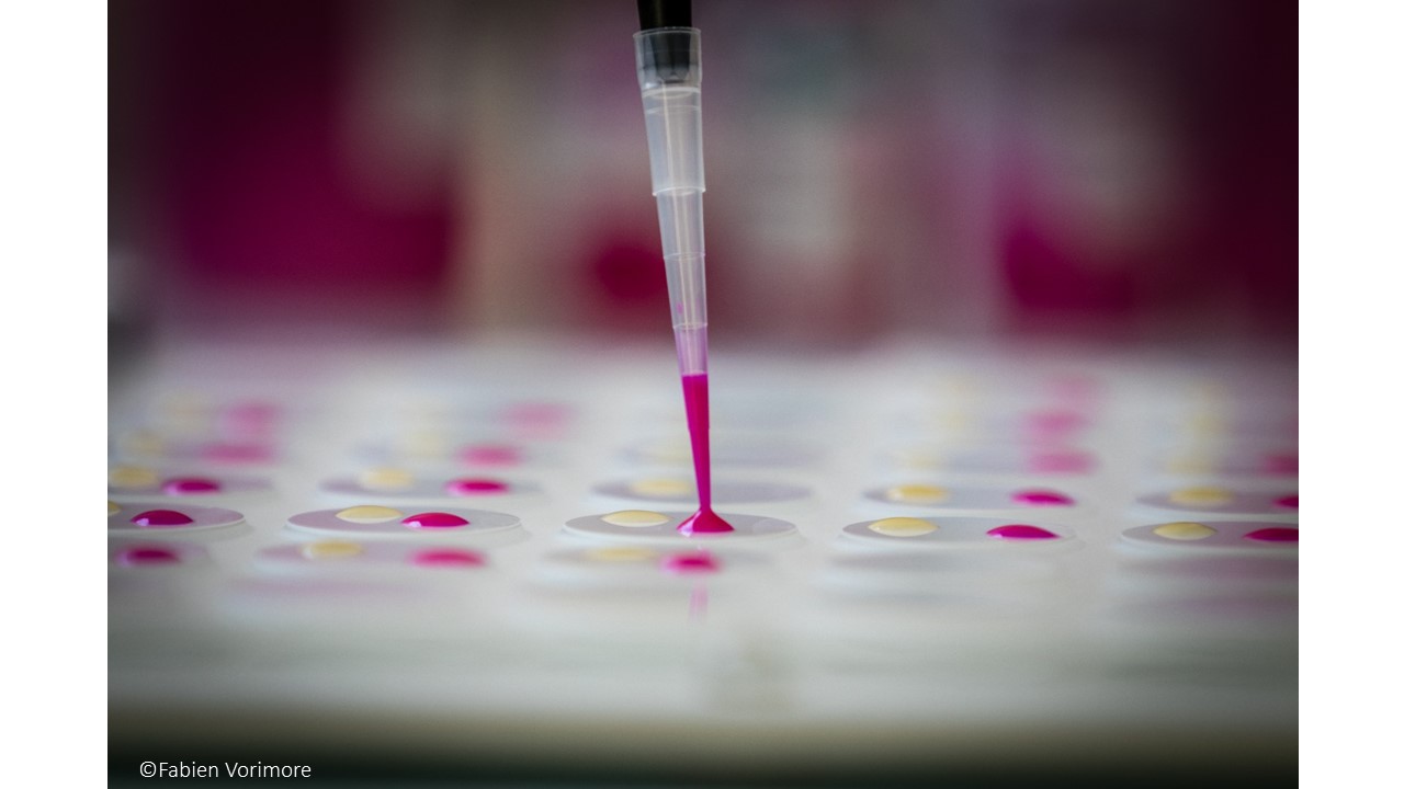
An ELISA is a plate-based laboratory technique making use of the binding between an antigen and its homologous antibody in order to identify and quantify the specific antigen or antibody in a sample. ELISAs are typically performed in 96-well (or 384-well) polystyrene plates, which will passively bind antibodies and proteins.

c-ELISA kits have to be previously validated according to EURL specifications. c-ELISA tests have to be performed according to manufacturer instructions.


INTRODUCTION

Antibodies also known as immunoglobulins (Ig) are gamma globulin proteins that are found in blood. These specialized immune proteins are produced following the introduction of an antigen into the body and possess the remarquable ability to combine with the antigen that triggered its production (specific affinity). The antibody recognises and bind to the antigenic determinant region of the antigen.

Enzyme-linked immunosorbent assays rely on specific antibodies to bind the target antigen, and a detection system to indicate the presence and quantity of antigen binding.


Brucella antigens (for review, Ducrotoy et al., 2016, DOI: 10.1016/j.vetimm.2016.02.002).

Like other gram negative bacteria, Brucella consist of cytoplasm surrounded by a cell envelope made of an inner membrane, periplasm and an outer membraine. The outer membrane contains two main classes of antigens: lipopolysaccharide (LPS) and detergent-soluble proteins (referred to as outer membrane proteins [Omp]). Structurally, the LPS can be either smooth (S) or rough (R) depending on the Brucella species. These two LPS differ in the presence in the former and absence in the latter of a long polysaccharide section, the O-polysaccharide


The antigens relevant to ruminant brucellosis can be classified into 2 categories: S-LPS and the related haptenic polysaccharides (O-polysaccharide linked with most core oligosaccharide sugars or core-O-polysaccharides ; the native haptens (NH) and the polysaccharide B) ; and protein antigens. The O-polysaccharide carries three basic antibody reactivities: A, M and C (Douglas and Palmer, 1988). In diagnosis, the criticial concept is the existence in the serum of an infected animal of a range of antibody reactivities (A>M, A= M and A<M) that in practice correspond to overlapping epitopes highly repeated in the O-polysaccharide (hence their colloective name as Brucella C epitope ; Weynants et al., 1997). Brucella O-polysaccharides cross-react with gram-negative bacteria that contain variable proportions of N-substituted perosamine. Among these bacteria, Yersinia enterocolitica serotype O:9 generates the strongest cross-reactivity.


Principle of C-ELISA (source: Laboratoryinfo.com)

This type of ELISA depends on the competitive reaction between the sample antigen and antigen bound to the wells of microtiter plate with the primary antibody.


1-First, the primary antibody is incubated with the sample. This results in the formation of Ag-Ab complex which are then added to the wells that have been coated with the same antigens.

2-After an incubation, unbound antibodies are washed off. The more antigen in the sample, more primary antibody will bind to the sample antigen. Therefore there will be smaller amount of primary antibody available to bind to the antigen coated on well.

3-Secondary antibody conjugated to an enzyme is added, followed by a substrate to elicit a chromogenic signal.


Concentration of color is inversely proportional to the amount of antigen present in the sample.


Before starting the work, read kit instruction carefully!


Results

The ELISA assay yields three different types of data output:

  1. Quantitative: ELISA data can be interpreted in comparison to a standard curve (a serial dilution of a known, purified antigen) in order to precisely calculate the concentrations of antigen in various samples.
  2. Qualitative: ELISAs can also be used to achieve a yes or no answer indicating whether a particular antigen is present in a sample, as compared to a blank well containing no antigen or an unrelated control antigen.
  3. Semi-Quantitative: ELISAs can be used to compare the relative levels of antigen in assay samples, since the intensity of signal will vary directly with antigen concentration.


Serological tests for Brucellosis [relayed from OIE Terrestrial Manual]:

The buffered Brucella antigen tests (rose bengal test and buffered plate agglutination test), the complement fixation test, the enzyme-linked immunosorbent assays (ELISA) or the fluorescence polarisation assay, are suitable tests for screening of herds/flocks and individual small ruminants, camelids and bovines (cattle and buffaloes). However, no single serological test is appropriate in each animal species and all epidemiological situations, and some of these tests are not adequate for diagnosing brucellosis in pigs. Therefore, the reactivity of samples that are positive in screening tests should be assessed using an established confirmatory or complementary strategy. The indirect ELISA or milk ring test performed on bulk milk samples is effective for screening and monitoring dairy cattle.


Several variations of the C-ELISA, using S-LPS or O-PS as antigens, have been described for cattle, small ruminants and pigs employing different antiglobulin-enzume conjugates, substrate or chromogens and antigens prepared from different smooth Brucella strains. Nevertheless, the technique used and the interpretation of results must have been validated in accordance with OIE requirements. It has been shown that the C-ELISA eliminates some but not all False Positive Serological Reactions (FPSR) caused by cross-reacting bacteria in cattle (Muñoz et al., 2005) and swine (Praud et al., 2012). In some cases, in ruminants or pigs, FSPR may be observed in C-ELISA while not in other S-LPS-based tests, including I-ELISA. Moreover, the C-ELISA reduces but not fully eliminates the reactions caused by antibodies produces in response to vaccination.

Some protocols are less sensitive or less specific than others; therefore results obtained from different assays are not always comparable. C-ELISA for diagnosing anti-Brucella antibodies in small ruminants and swine is essentially te same as that described for cattle, but the cut-off should have been properly establhised for these species using the appropriate validation techniqes.


Whatever the C-ELISA format used:

i) a positive and a negative control are included in each plate. Optical Density (OD) ranges to be obtained with these two controls must be established to define the criteria for validating each plate results. The OD of the positive control is the one with which the OD of each test serum is compared to establish the final result -negative of positive).

ii) an additional positive serum (internal control) must be included in each plate to validate the repeatability of the test from plate to plate and from day to day (control chart).


Complement Fixation Test

CFT

The  complement fixation test (CFT) is used to detect the presence of specific antibodies in the patient’s serum. This test is based on the use of complement, a Biologically labile serum factor that causes the immune cytolysis i.e. lysis of antibody coated cells.


Principle (source Laboratoryinfo.com)

It is the nature of the complement to be activated when there is formation of antigen-antibody complex.
The first step is to heat the serum at 56°C to destroy patient’s complement.

A measured amount of complement and antigen are then added to the serum.

If there is presence of antibody in the serum, the complement is fixed due to the formation of Ag-Ab complex. If no antibody is present then the complement remains free.

To determine whether the complement has been fixed, sheep RBCs and antibodies against sheep RBCs are added.

Interpretation

Positive test : The available complement is fixed by Ag-Ab complex and no hemolysis of sheep RBCs occurs. So the test is positive for presence of antibodies.

Negative test : No Ag-Ab reaction occurs and the complement is free. This free complement binds to the complex of sheep RBC and it’s antibody to cause hemolysis, causing the development of pink color.


Indirect Enzyme Linked Immunosorbent Assay

I-ELISA

An ELISA is a plate-based laboratory technique making use of the binding between an antigen and its homologous antibody in order to identify and quantify the specific antigen or antibody in a sample. ELISAs are typically performed in 96-well (or 384-well) polystyrene plates, which will passively bind antibodies and proteins.

i-ELISA kits have to be validated according to EURL spectifications. i-ELISA tests have to be performed according to manufacturer instructions.


INTRODUCTION

Antibodies also known as immunoglobulins (Ig) are gamma globulin proteins that are found in blood. These specialized immune proteins are produced following the introduction of an antigen into the body and possess the remarquable ability to combine with the antigen that triggered its production (specific affinity). The antibody recognises and bind to the antigenic determinant region of the antigen.

Enzyme-linked immunosorbent assays rely on specific antibodies to bind the target antigen, and a detection system to indicate the presence and quantity of antigen binding.


Brucella antigens (for review, Ducrotoy et al., 2016, DOI: 10.1016/j.vetimm.2016.02.002).

Like other gram negative bacteria, Brucella consist of cytoplasm surrounded by a cell envelope made of an inner membrane, periplasm and an outer membraine. The outer membrane contains two main classes of antigens: lipopolysaccharide (LPS) and detergent-soluble proteins (referred to as outer membrane proteins [Omp]). Structurally, the LPS can be either smooth (S) or rough (R) depending on the Brucella species. These two LPS differ in the presence in the former and absence in the latter of a long polysaccharide section, the O-polysaccharide.


The antigens relevant to ruminant brucellosis can be classified into 2 categories: S-LPS and the related haptenic polysaccharides (O-polysaccharide linked with most core oligosaccharide sugars or core-O-polysaccharides ; the native haptens (NH) and the polysaccharide B) ; and protein antigens. The O-polysaccharide (O-PS) carries three basic antibody reactivities: A, M and C (Douglas and Palmer, 1988). In diagnosis, the criticial concept is the existence in the serum of an infected animal of a range of antibody reactivities (A>M, A= M and A<M) that in practice correspond to overlapping epitopes highly repeated in the O-polysaccharide (hence their colloective name as Brucella C epitope ; Weynants et al., 1997).  Brucella O-polysaccharides cross-react with gram-negative bacteria that contain variable proportions of N-substituted perosamine. Among these bacteria, Yersinia enterocolitica serotype O:9 generates the strongest cross-reactivity.


Principle of I-ELISA (source: Laboratoryinfo.com)

1-Coating: the protein antigen to be tested for is added to each well of ELISA plate, where it is given time to adhere to the plastic through charge interactions.

2-Blocking: a solution of non-reacting protein is added to block any plastic surface in the well that remains uncoated by the protein antigen.

3-Then the serum is added, which contains a mixture of the serum antibodies, of unknown concentration, some of which may bind specifically to the test antigen that is coating the well.

4-Detection: afterwards, a secondary enzyme-conjugated antibody is added, which will bind to the antibody bound to the test antigen in the well.  The secondary antibody is usually an anti-species antibody and is often polyclonal. A number of enzymes have been employed for ELISA, including alkaline phosphatase, horseradish peroxidase, and B-galactosidase.

5-Reading results: a substrate for this enzyme is then added. Often, this substrate changes colour upon reaction with the enzyme. The colour change shows that secondary antibody has bound to primary antibody, which strongly implies that the donor has had an immune reaction to the test antigen.

The higher the concentration of the primary antibody that was present in the serum, the stronger the colour change. Often a spectrometer is used to give quantitative values for colour strength.

Before starting the work, read kit instruction carefully!


Results

The ELISA assay yields three different types of data output:

  1. Quantitative: ELISA data can be interpreted in comparison to a standard curve (a serial dilution of a known, purified antigen) in order to precisely calculate the concentrations of antigen in various samples.
  2. Qualitative: ELISAs can also be used to achieve a yes or no answer indicating whether a particular antigen is present in a sample, as compared to a blank well containing no antigen or an unrelated control antigen.
  3. Semi-Quantitative: ELISAs can be used to compare the relative levels of antigen in assay samples, since the intensity of signal will vary directly with antigen concentration.


Serological tests for Brucellosis [relayed from OIE Terrestrial Manuel]:

The buffered Brucella antigen tests (BBATs; rose bengal test and buffered plate agglutination test), the complement fixation test, the enzyme-linked immunosorbent assays (ELISA) or the fluorescence polarisation assay, are suitable tests for screening of herds/flocks and individual small ruminants, camelids and bovines (cattle and buffaloes). However, no single serological test is appropriate in each animal species and all epidemiological situations, and some of these tests are not adequate for diagnosing brucellosis in pigs. Therefore, the reactivity of samples that are positive in screening tests should be assessed using an established confirmatory or complementary strategy. The indirect ELISA or milk ring test performed on bulk milk samples is effective for screening and monitoring dairy cattle.


The I-ELISAs that use S-LPS or O-PS as antigens are highly sensitive for the detection of anti-Brucella antibodies in cattle, small ruminants and pigs, but are not capable of fully resolving the problem of differentiating between antibodies resulting from B. abortus S19 and B. melitensis Rev.1 vaccination. The B. abortus RB51 may also interfere in S-LPS-based I-ELISAs. Using I-ELISA standardised against the OIE standard sera, the diagnostic sensitivity should be equal or greater than that of the BBATs or the CFT (complement fixation test) in the testing of infected cattle, small ruminants and pigs. However, the specificity should be lower (Praud et al., 2012). The problem of False Positive Serological Reactions (FPSR) may be reduced but not fully resolved, in pigs in particular, by performing I-ELISAs using extracts from rough strains of Brucella. Most FPSR are a result of cross reaction with the O-PS portion of the S-LPS molecule; cross-reaction amoung core epitopes is less frequent but does exist.


Several commercial I-ELISAs are available. Some protocols are less sensitive or less specific than others; therefore results obtained from different assays are not always comparable. I-ELISA for diagnosing anti-Brucella antibodies in small ruminants and pigs is essentially the same as that described for cattle, but the cut-off should have been properly established for these species using the appropriate validation techniques, and moreover, I-ELISA for sheep and goats should be standardised against the ISaBmS (McGiven et al., 2011).


Whatever the I-ELISA format used:

i) a positive and a negative control are included in each plate. Optical Density (OD) ranges to be obtained with these two controls must be established to define the criteria for validating each plate results. The OD of the positive control is the one with which the OD of each test serum is compared to establish the final result -negative of positive).

ii) an additional positive serum (internal control) must be included in each plate to validate the repeatability of the test from plate to plate and from day to day (control chart).


Successfull european contributions to the Brucellosis network in Chicago !

72nd Annual Brucellosis Research Conference

The International Brucellosis Society organized its annual 2-day conference  in Chicago on November 2 and 3, 2019, bringing together 120 participants from 27 countries and including 18 students. In total, the conference counted 33 oral communications and 21 posters. The European and French teams were well represented with 12 oral presentations and 2 posters.

The program scanned the different aspects of the research, including cellular interactions between Brucella and host cells (with a guest lecture by Suzana Salcedo, CNRS UMR 5086, University of Lyon), experimental results on new vaccine approaches and several communications related to particular human cases and Brucella excretion in milk and cheese.


Seven European countries contributed actively to the conference programme : ANSES (EURL for Brucellosis, France), APHA (UK), BfR (Germany), CITA (Spain), CNRS (France), INSERM (France), ISZAM (Italy) and NARILIS (Belgium). The EURL team presented 2 oral communications and 1 poster, by Guillaume Girault, Claire Ponsart and Luca Freddi respectively.

Noteworthy was an oral presentation on the STAR-IDAZ project, with Brucellosis as a priority topics. More information will be given in our next newsletter.


Next conference will be organised by ISZAM in Italy, 2020, September 29th to October, 2nd. A great opportunity for EU teams to meet and present their research and results, save the date!

Pages